Home
/ Posterior Neck Muscle Diagram : Pin em Anatomy Notes - Other branches of the cervical plexus innervate the vertebral muscles, and provide cutaneous innervation to parts of the neck and scalp.
Posterior Neck Muscle Diagram : Pin em Anatomy Notes - Other branches of the cervical plexus innervate the vertebral muscles, and provide cutaneous innervation to parts of the neck and scalp.
Posterior Neck Muscle Diagram : Pin em Anatomy Notes - Other branches of the cervical plexus innervate the vertebral muscles, and provide cutaneous innervation to parts of the neck and scalp.. The transverse cervical and suprascapularveins also lie in the posterior triangle the subclavian, transverse cervical and suprascapular veins are accompanied by their respective arteries in the posterior triangle. The posterior triangle of the neck contains many muscles, which make up the borders and the floor of the area. See full list on teachmeanatomy.info The external jugular vein is one of the major veins of the neck region. Jan 20, 2018 · neck muscles are bodies of tissue that produce motion in the neck when stimulated.
What muscles are on the back of the neck? The cervical plexus forms within the muscles of the floor of the posterior triangle. They consist of 3 main groups of muscles: These muscles are mainly responsible for the movement of the head in all directions. The subclavian vein is often used as a point of access to the venous system, via a central catheter.
The Posterior Triangle of the Neck - dummies from www.dummies.com The external jugular vein is one of the major veins of the neck region. The subclavian vein is often used as a point of access to the venous system, via a central catheter. The muscles of the neck run from the base of the skull to the upper back and work together to bend the head and. Other branches of the cervical plexus innervate the vertebral muscles, and provide cutaneous innervation to parts of the neck and scalp. Feb 17, 2015 · superficial muscles are the muscles closest to the skin surface and can usually be seen while a body is performing actions. Learn vocabulary, terms, and more with flashcards, games, and other study tools. Jun 17, 2021 · muscles of the neck (musculi cervicales) the muscles of the neck are muscles that cover the area of the neck. See full list on teachmeanatomy.info
What are the deep muscles of the neck?
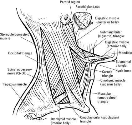
The cervical plexus forms within the muscles of the floor of the posterior triangle. Within the posterior triangle, the external jugular vein pierces the investing layer of fascia and empties into thesubclavian vein. Anterior, middle and posterior scalenes Start studying neck muscles (posterior). It lies relatively superficialin the posterior triangle, leaving it vulnerable to injury. It crosses the posterior triangle in an oblique, inferoposterior direction, within the investing layer of fascia. Neck_muscle_anatomy_model 3/4 neck muscle anatomy model perform extraoral and intraoral patient examinations. See full list on teachmeanatomy.info Learning objectives open each chapter with goals to be accomplished, and serve as checkpoints for comprehension, skills mastery, and exam preparedness. Start studying posterior neck muscles. Jun 17, 2021 · muscles of the neck (musculi cervicales) the muscles of the neck are muscles that cover the area of the neck. The trunks of the brachial plexusalso cross the floor of the posterior triangle. The distal part of the subclavian arterycan be located as it emerges between the anterior and middlescalenemuscles.
Anterior, lateral and posterior groups, based on their position in the neck. The subclavian vein is often used as a point of access to the venous system, via a central catheter. Many in the neck help to stabilize or move the head. The muscle then crosses underneath the scm to enter the anterior triangleof the neck. Jun 17, 2021 · muscles of the neck (musculi cervicales) the muscles of the neck are muscles that cover the area of the neck.
Anterior Triangle of Neck - Submental and Muscular ... from i1.wp.com See full list on teachmeanatomy.info The external jugular vein is one of the major veins of the neck region. These muscles are mainly responsible for the movement of the head in all directions. It lies relatively superficialin the posterior triangle, leaving it vulnerable to injury. As it crosses the first rib, it becomes the axillaryartery, which goes onto supply the upper limb. The muscles of the neck run from the base of the skull to the upper back and work together to bend the head and. Learning objectives open each chapter with goals to be accomplished, and serve as checkpoints for comprehension, skills mastery, and exam preparedness. It is split into two bellies by a tendon.
Start studying posterior neck muscles.
It crosses the posterior triangle in an oblique, inferoposterior direction, within the investing layer of fascia. The muscle then crosses underneath the scm to enter the anterior triangleof the neck. Learning objectives open each chapter with goals to be accomplished, and serve as checkpoints for comprehension, skills mastery, and exam preparedness. A significant muscle in the posterior triangle region is the omohyoid muscle. The cervical plexus forms within the muscles of the floor of the posterior triangle. It is split into two bellies by a tendon. The trunks of the brachial plexusalso cross the floor of the posterior triangle. What muscles are on the back of the neck? The posterior neck triangle is covered superficially to deep by the skin superficial and deep cervical fascia and the platysma muscle. The accessory nerve (cn xi) exits the cranial cavity, descends down the neck, innervates sternocleidomastoid and enters the posterior triangle. It descends down the neck, within the prevertebralfascia, to innervate the diaphragm. A number of vertebral muscles (covered by prevertebral fascia) form the floorof the posterior triangle: Formed by the retromandibular and posterior auricular veins, it lies superficially, entering the posterior triangle after crossing the sternocleidomastoid muscle.
Learning objectives open each chapter with goals to be accomplished, and serve as checkpoints for comprehension, skills mastery, and exam preparedness. A significant muscle in the posterior triangle region is the omohyoid muscle. It is split into two bellies by a tendon. A number of vertebral muscles (covered by prevertebral fascia) form the floorof the posterior triangle: The muscles of the neck run from the base of the skull to the upper back and work together to bend the head and.
Anatomy of the Head and Neck - Medical Illustrations ... from www.medical-artist.com The muscle then crosses underneath the scm to enter the anterior triangleof the neck. Other branches of the cervical plexus innervate the vertebral muscles, and provide cutaneous innervation to parts of the neck and scalp. The accessory nerve (cn xi) exits the cranial cavity, descends down the neck, innervates sternocleidomastoid and enters the posterior triangle. Jun 17, 2021 · muscles of the neck (musculi cervicales) the muscles of the neck are muscles that cover the area of the neck. It crosses the posterior triangle in an oblique, inferoposterior direction, within the investing layer of fascia. They consist of 3 main groups of muscles: The posterior triangle is crossed about 25 cm above the clavicle by the inferior belly of the omohyoid muscle which divides the space into two triangles. The distal part of the subclavian arterycan be located as it emerges between the anterior and middlescalenemuscles.
The muscles of the neck run from the base of the skull to the upper back and work together to bend the head and.
Learn vocabulary, terms, and more with flashcards, games, and other study tools. Start studying neck muscles (posterior). The external jugular vein is one of the major veins of the neck region. See full list on teachmeanatomy.info Other branches of the cervical plexus innervate the vertebral muscles, and provide cutaneous innervation to parts of the neck and scalp. Neck_muscle_anatomy_model 3/4 neck muscle anatomy model perform extraoral and intraoral patient examinations. The muscle then crosses underneath the scm to enter the anterior triangleof the neck. The subclavian vein is often used as a point of access to the venous system, via a central catheter. What are the deep muscles of the neck? Formed by the retromandibular and posterior auricular veins, it lies superficially, entering the posterior triangle after crossing the sternocleidomastoid muscle. Start studying posterior neck muscles. More images for posterior neck muscle diagram » The accessory nerve (cn xi) exits the cranial cavity, descends down the neck, innervates sternocleidomastoid and enters the posterior triangle.
The muscle then crosses underneath the scm to enter the anterior triangleof the neck neck muscle diagram. It lies relatively superficialin the posterior triangle, leaving it vulnerable to injury.